PBL 3 Fractures and dislocations
Clinical Outcomes
Discuss the basic epidemiology, presentation, investigation, management, complications and prognosis of fractures - with focus on the main fracture presentations including hip/femur, knee, shoulder, wrist, scaphoid, tibia and ankle
Ankle fractures (1)
- Commonly a low-energy fracture of either the lateral and/or medial malleolus
- Includes fracture of the posterior malleolus
- Vascular compromise is rare
-
Over 65/older women 1.5-5.3 per 1000 people
-
Highest frequency is white women and lowest being non-white men
-
Proper immobilisation, analgesia and timely orthopaedic referral can greatly enhance patient comfort and ensure optimal outcome
-
Displaced fractures commonly treated with an open reduction and internal fixation
-
Open fractures (bone pierces skin) require emergency treatment by surgeons
-
Diagnostic factors: Recent trauma, ankle pain and swelling, inability to weight-bear
-
Other factors: Ankle deformity, crepitus (joint movement sounds), tenderness of the proximal fibula
-
Risk factors: Low-energy fall, osteoporosis, multiple falls, inversion injury to ankle
-
Ottawa ankle rules: Help decide when to order x-rays, if there is posterior lateral or medial bony tenderness within 6cm of the distal aspect of the fibula or tibia or inability to weight bear 4 steps at the scene then ankle x-ray should be ordered.
First investigation to order is a plain x-ray
Hip fracture (2)
-
Occur predominantly in older people as risk increases significantly with age
-
In younger patients, they usually result from higher energy trauma
-
Associated commonly with low-energy injury (e.g. a fall) and osteoporosis
-
Treatment is most commonly surgical
-
Any fracture of the femur distal to the femoral head and proximal to a level a few cm below the lesser trochanter
-
76'000 hip fractures each year
-
Occupy 4'000 NHS beds at any one time
-
History of fall/trauma
-
Pain in the affect hip, groin or thigh
-
Inability to bear weight or move hip
-
Shortened, externally rotated leg
-
Risk factors: Osteoporosis/osteopenia, old age, falls, low BMI
First investigations to order are plain radiographs, full blood count, urea and electrolytes and glucose
Wrist fracture (3)
-
Non-displaced fractures do not have any deformity and are treated with immobilisation in a splint or cast
-
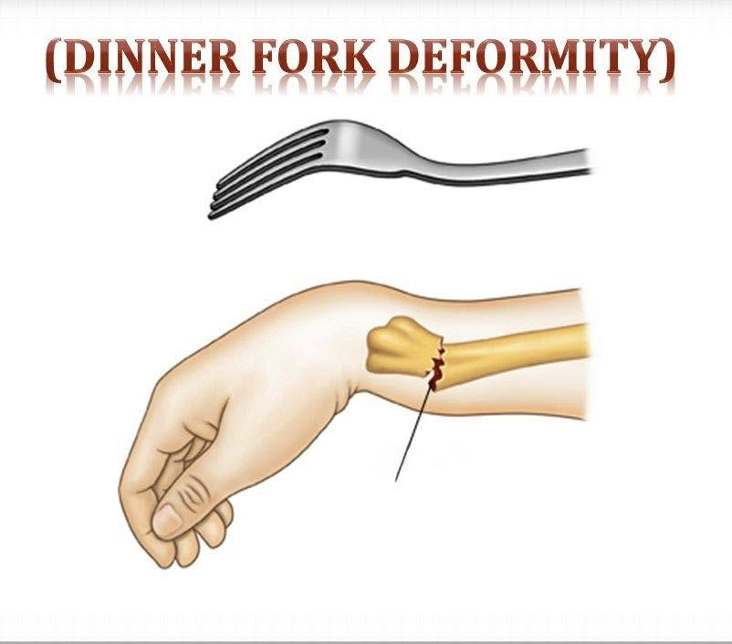
Displaced fractures present with a 'dinner fork' deformity and require closed reduction and possible surgical fixation
-
Fractures that affect the distal ends of the radius, ulna and carpus. Distal radius are one of the most common injuries
-
Incidence of distal radius fractures is 36.8/10'000 in women and 9.0/10'000 in men over 35
-
Women of any age - 170.2/100'000
-
Men of any age - 83.9/100'000
-
Presence of risk factors
-
Wrist pain
-
Tenderness over the distal radius
-
Swelling
-
Deformity - 'dinner fork'
-
Risk factors: Trauma, osteopenia, osteoporosis
First investigation to order is plain x-rays of the wrist
Long bone fracture
-
Long bones defined as humerus, radius, ulna, femur, tibia and fibula
-
At least 2 90
orthogonal x-rays (sagittal, transverse, coronal) should be obtained with inclusion of joints proximal and distal to site -
Incidence of fractures in Britain is about 3.6%, 1.8M fractures in England each year
-
Tibia is most frequently fractured, 6.6% of all fractures
-
Pain
-
Soft-tissue swelling
-
Ecchymosis - bruise
-
Altered nerve sensation
-
Impaired motor function
-
Risk Factors: Direct/indirect trauma, osteoporosis and chronic renal failure
First investigations to order are x-ray limb, full blood count blood typing and cross-matching
Describe the radiological principles behind diagnosing and describing fractures
Ottawa ankle rules (1)
- Help decide when to order x-rays
- Ankle x-rays should be ordered if there is posterior lateral or medial bony tenderness within 6cm of the distal aspect of the fibula or tibia or inability to weight bear 4 steps
- Ankle fractures are diagnoses mainly from plain x-rays in orthogonal planes
Hip fractures (2)
- Order plain radiographs with both sagittal and coronal views
- Radiography is the definitive test to confirm the diagnosis
- If radiograph does not show a fracture but strong suspicion of one, request MRI
- Classify fractures radiographically into intracapsular (in joint capsule) or extracapsular (out of join capsule) to determine surgical managemetn
Wrist fractures (3)
- Order plain x-rays in all patients
- Order MRI if there is suspected scaphoid fracture
Long bone fractures (4)
- Order plain x-rays in all patients with suspected lower limb fracture
Discuss the basic epidemiology, presentation, investigation and management of compartment syndrome
Compartment syndrome of extremities (5)
-
Incidence of acute compartment syndrome is difficult to ascertain because concrete diagnostic criteria are elusive
-
0.7 per 100'000 women, 7.3 per 100'000 men
-
Presence of risk factors (fractures, osteoporosis)
-
Pain
-
Pressure (muscle tightness)
-
Paraesthesia - abnormal skin sensation
-
Paralysis
-
Risk factors: Trauma, bleeding disorder, compression support and thermal injury
First investigation to order is compartment pressure measurement
- Significant muscle necrosis - amputation may need to be considered
- Casts or dressings should be split completely
Describe the common joint dislocations and their complications and management - with focus on the main dislocation presentations including shoulder, elbow, hip, knee and ankle
Joint dislocation (6)
-
Complete separation of 2 articulating bony surfaces
-
Common sites: shoulder, finger, patella, elbow and hip
-
Partial/incomplete dislocation - subluxation
-
Glenohumeral joint is the most mobile joint - vulnerable to dislocation

-
Anterior shoulder dislocations account for more than 95% of shoulder dislocations and the most common discloation
-
Incidence has bi-modal peaks: men between adolescence and 30 years and women aged 61-80
-
23.9 per 100'000 people in general population
-
Presence of risk factors
-
Characteristic posturing of joint
-
Pain
-
Inability to move joint
First investigations to order are plain x-rays of suspected dislocated site
Discuss the methods of imaging bone and the advantages and disadvantages of each
X-ray (11)
- Fly through many kinds of matter
- X-rays collide with electrons: sometimes transfers all its energy and gets absorbed, or transfers some energy and the rest scatters
- Frequency of outcomes depends on the number of electrons hit
- Collisions more likely for dense material or elements of higher atomic number
- Bones are dense - absorb x rays well
- Soft tissue are less dense - absorb less, is penetrated
Magnetic Resonance Imaging (MRI) (7)
- MRIs use protons which is abundant in the human body
- Protons spin which creates a small magnetic charge
- When a strong magnetic field is introduced the protons align with the field
- Radiofrequency pulse is introduced which disrupts the protons into a 90
or 180 realignment - When radiofrequency is turned off the protons realign with the field
- This releases electromagnetic energy and the MRI detects this energy and differentiates between tissues based on the quickness of release
Computer Tomography (CT) (10)
- Creates cross-sectional images with the help of computer processing
- CT uses a motorised x-ray source that shoots narrow beams of x-rays as it rotates
- Digital x-ray detectors opposite the x-ray source which transmit to a computer
- Images individually are 2D but stacking them together can create a 3D image
- Computers use iterative reconstruction which uses forward projections detected by the CT to create an image
- Python code tutorial
Positron Emission Tomography (PET) (8)
- Tiny tracers with specifically designed radioactive molecules
- PET tracks the position of trackers
- Reveals activity in 3D
- Tracer can be any molecule (sugar, protein, hormone) each are tailored to a specific function in the body
- Radioactive atom on tracer loses radioactivity which releases a positron
- Positron + Electron they combine and destroy each other which releases energy as two gamma rays that travel out and in opposite directions
- PET scanner is like a geiger counter (radioactivity detector)
- This detects the two gamma rays and can calculate where the tracer must be
Functional MRI (fMRI) (9)
- Images of activity
- Observation of the response of protons
- Determine properties of the tissue the signal came from
- Differences in the magnetic properties of oxygenated vs deoxygenated blood
Ultrasound
- High frequency sound waves emitted from a probe
- Sound waves bounce back off tissues - echo
- Echoes detected and translated to image
- No radiation
Advantages/disadvantages
| Method | Advantage | Disadvantage |
|---|---|---|
| X-ray | See bones, no need for tracers, quicker | Can't see tissue, only one image angle produced |
| MRI | See tissues, no need for tracers | Slow, high cost, loud, risk for people with metal on self |
| CT | See bones and tissue, no need for tracers, | Intensive computational power needed |
| PET | See activity of tissues | Needs tracers |
| fMRI | See activity of tissues | Slow, imprecision (regions rather than individual tssues) |
| Ultrasound | See tissues, no radiation | Only tissues |
Discuss the indication for prescribing Paracetamol
Indication (12)
- Mild to moderate pain
- Pyrexia (fever)
- Acute migraine
Discuss the efficacy, safety, suitability and routes of administration for Paracetamol
Routes of administration (12)
- Mouth
- Intravenous infusion
- Rectum
Safety information
- No evidence that taking paracetamol during pregnancy causes autism in children
- Multiple incidents of overdose for adult inpatients with low body-weight (<50kg)
Side-effects
- Thrombocytopenia (low platelet count)
In Lakia's case she was given paracetamol for the pain
- 0.5-1g every 4-6 hours. Maxes out at 4g for a day
In Billy-Joe's case he was given paracetamol for the pain - 480-500mg every 4-6 hours. Maxes out at 4 doses a day
Quiz
- X-ray - bones
- CT - bones and tissues
- MRI - tissues
- Ultrasound - tissues
- Nerve damage
- Blood vessel damage
- Bone fragments piercing skin (compound fracture)
- Infection
- Displaced means the fracture causes one of the fractured bones to not align with the other
- Realign the bone, surgery is needed as splints/casts cannot realign by themselves
- Very mobile joints
Reference/s
- bestpractice.bmj.com. (n.d.). Ankle fractures - Symptoms, diagnosis and treatment | BMJ Best Practice. [online] Available at: https://bestpractice.bmj.com/topics/en-gb/385?q=Ankle fractures&c=suggested [Accessed 2 Oct. 2026].
- MFS (2026). Hip fractures - Symptoms, diagnosis and treatment | BMJ Best Practice US. [online] Bmj.com. Available at: https://bestpractice.bmj.com/topics/en-gb/387 [Accessed 2 Oct. 2026].
- Bmj.com. (2025). Wrist fractures - Symptoms, diagnosis and treatment | BMJ Best Practice US. [online] Available at: https://bestpractice.bmj.com/topics/en-gb/392 [Accessed 2 Oct. 2026].
- Bmj.com. (2025). Long bone fracture - Symptoms, diagnosis and treatment | BMJ Best Practice US. [online] Available at: https://bestpractice.bmj.com/topics/en-gb/386 [Accessed 2 Oct. 2026].
- bestpractice.bmj.com. (n.d.). Compartment syndrome of extremities - Symptoms, diagnosis and treatment | BMJ Best Practice. [online] Available at: https://bestpractice.bmj.com/topics/en-gb/502 [Accessed 2 Oct. 2026].
- Bmj.com. (2026). Joint dislocation - Symptoms, diagnosis and treatment | BMJ Best Practice US. [online] Available at: https://bestpractice.bmj.com/topics/en-gb/583?q=Joint dislocation&c=suggested [Accessed 2 Oct. 2026].
- NIBIB (2013). How Does an MRI Scan Work? Available at: https://www.youtube.com/watch?v=1CGzk-nV06g [Accessed 3 Oct. 2026].
- Imperial College London (2016). How does a PET scan work? YouTube. Available at: https://www.youtube.com/watch?v=yrTy03O0gWw [Accessed 3 Oct. 2026].
- Neuroscientifically Challenged (2019). 2-Minute Neuroscience: Functional Magnetic Resonance Imaging (fMRI). YouTube. Available at: https://www.youtube.com/watch?v=rJjHjnzmvDI [Accessed 3 Oct. 2026].
- NIBIB gov (2017). How Does a CT Scan Work? YouTube. Available at: https://www.youtube.com/watch?v=l9swbAtRRbg [Accessed 3 Oct. 2026].
- TED-Ed (2015). How X-rays see through your skin - Ge Wang. Available at: https://www.youtube.com/watch?v=gsV7SJDDCY4 [Accessed 3 Oct. 2026].
- British National Formulary (2025). Paracetamol. [online] NICE. Available at: https://bnf.nice.org.uk/drugs/paracetamol/ [Accessed 3 Oct. 2026].