Functional Histology
LO: Explain the organisation and structure of the four basic human tissue types (epithelium, connective tissue, muscle and nervous tissue) and describe how cell and tissue structure facilitates normal organ function
Histology - Study of tissues.
Tissue:
- Nervous - Ectoderm
- Epithelial - Endoderm
- Muscle - Mesoderm
- Connective - Mesoderm
Epithelium:

Apical - Top
Basal - Bottom
flowchart TD
	A[Epithelial Tissues] --> B{Layers of cells?}
	B --> Simple
	B --> Compound
	Simple --> C{Cell Shapes?}
	Compound --> D{Change Shapes?}
	C --> Pseudostratified
	C --> Squamous
	C --> Cuboidal
	C --> Columnar
	D --> Stratified
	D --> Transitional
	Pseudostratified --> E{Forming Cilia?}
	E --> Ciliated
	E --> Non-Ciliated
	Stratified --> F{Cell Shapes?}
	F --> G[Stratified squamous]
	F --> H[stratified cuboidal]
	F --> I[stratified columnar]
	G --> J{Containing keratin?}
	J --> Keratinised
	J --> Non-keratinised
This flow chart took me forever holy

Simple - Just single layer
Stratified - Tall, a few layers
Squamous - Deflated very flat
Cuboidal - Very cube not flat
Columnar - Rectangle cigarettes
Pseudostratified - Very tall but still single layer
Cilia - Absorption and secretion, microvilli and goblet cells to increase surface area
Epithelial tissue
flowchart TD
	Epithelial --> A[Covering Epithelium]
	Epithelial --> B[Glandular Epithelium]
Exocrine - Uses a duct to secrete
Endocrine - No duct, secrete directly into blood
Epithelial
- Ectoderm, Mesoderm, Endoderm
- Line and cover surfaces
- Cells join via adhesion molecules and junctional complexes
- Anchor to basement membrane
- Cells have structural and functional polarity
Simple Epithelium - Single layer of cells so good for gas exchange (lungs)
flowchart LR
	A[More cytoplasm] --> B[More organelle]
	B --> C[More secretion]
Transitional
- Extend and return
- Different shapes
- Changes shape
Muscle
- Skeletal - Multiple nuclei, Striated
- Cardiac - Single nucleus, Striated, Branched
- Smooth - Single nucleus, non-striated
| Nucleus | Striated | Branched | |
|---|---|---|---|
| Skeletal | Multiple | ✅ | |
| Cardiac | One | ✅ | ✅ |
| Smooth | One |
Skeletal
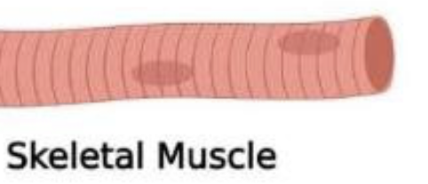
- Actin and Myosin, leads to sarcomeres so striated
Smooth
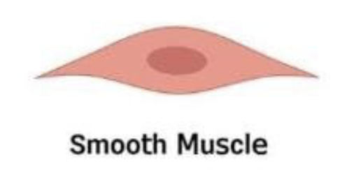
- Spindle shape
- Tapered edge
- Thick centre
Cardiac
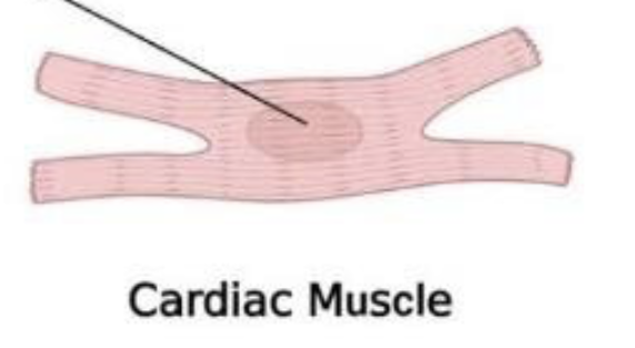
- Short, branched
- Striated
- Intercalated discs where muscles connect at the branched bit.
Connective Tissue
- Cells
- Fiber
- Ground substance (gel)
flowchart TD
	A[Connective Tissue] --> B[Connective Tissue Proper]
	A --> C[Supporting Connective Tissue]
	A --> D[Fluid Connective Tissue]
	B --> Loose
	B --> Dense
	C --> Cartilage
	C --> Bone
	D --> Blood
	D --> Lymph
Proper
- Hold organs
- Loose, more gel less fiber
- Dense, less gel more fiber
Blood
- Erythroid precursors -> Red blood cells
- Megakaryocyte -> Platelets
- Neutrophils look like Mickey Mouse
- Macrophage means big eat
- Plasma cells have eccentric nucleus (on one side)
Nervous
- Schwann cells (neurolemmocytes) - forms myelin sheath
- Endoneurium - thin layer of connective tissue