Embryology
LO: Describe the key stages and timelines of embryological, foetal and neonatal development
Embryology
- Branch of biology - development of organisms
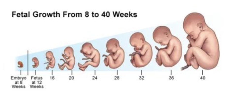
- Embryonic development in humans covers the first 8 weeks of development after fertilisation; at the start of the 9th week the embryo is termed a foetus
- Normal period of gestation (pregnancy) is ~9 months or 40 weeks
- Assists women's reproductive health
Embryological development
| Week | Stage |
|---|---|
| 1 | Fertilisation to implantation |
| 2 | Bilaminar germ disc, Amniotic cavity and primary yolk sac |
| 3 | Gastrulation: Trilaminar germ disc, primitive streak, notochord, neural tube and body axes/growth of embryonic disc |
| 3-8 | Organogenesis |
| 8-birth | Foetal period: Maturation tissues and organs and rapid growth of the foetal body |
Foetal development
Week 1
- Contact of the spermatozoon and ovum, normally in the ampulla (12-24 hrs after ovulation)
- Fusion of female and male pronucleus (2 haploid to form a diploid zygote)
- Two-cell zygote - cell cleavage into morula
- Ingress of fluid forms a blastocyst cavity (~4 days after fertilisation)
- Blastocyst consists of 2 cell layers: inner cell mass - embryoblast, outer cell mass - trophoblast
- Uterine secretory phase: Endometrium wall forms nutrient rich bed that receives and nourishes the blastocyst via diffusion
LO: Describe the development of the bilaminar germ disc
Week 2
- Blastocyst splits into two layers to form the bilaminar germ disc
Epiblast - high columnar cells, Hypoblast - cuboidal cells - Epiblast cells is the source of all embryonic tissues
- Formation of amniotic cavity and primary yolk sac
- Cytotrophoblast and syncyntiotrophoblast form
- Formation of chorionic cavity and plate
- Development of uteroplacental circulation
- Connecting stalk (primitive umbilical cord)
- Formation of the secondary yolk sac
LO: Describe the development of the 3 germ layers and what they develop into
Week 3
- Diffusion can no longer supply nutrients needed to develop embryo
- Formation of primitive blood vessels connected with the placenta
- Formation of the primitive streak, pit and node - linear groove in the epiblast cells of bilaminar layer
- Proliferation and migration of epiblast cells into primitive streak give rise to the trilaminar germ disc through a process of invagination or ingression
- Formation of three cell germ layers: ectoderm, mesoderm, endoderm
- Process of invagination of some epiblast mesenchyme cells
- Replace hypoblast to become endoderm
- Mesoderm formed in between
- Epiblast cells remaining on surface are the ectoderm
Gastrulation
- Formation of the notochord
- Mesenchyme cells migrate from primitive pit cranially under ectoderm to the oropharyngeal membrane
- Induces the overlying ectoderm to form of neural plate
- Sidedness determination
- Establishment of the body axes
- Differentiation of mesodermal elements
- Paraxial mesoderm
- Intermediate mesoderm
- Lateral mesoderm
Trilaminar germ disc
- Ectoderm: Nervous system, epidermis, some glands, sensory epithelium
- Mesoderm: Lining of body cavity and surround organs, muscle, connective tissue, bone, blood and blood vessels
- Endoderm: Internal lining of body systems
Ecto - outside, Meso - middle, Endo - inside, Derm - skin
- All three germ layers originate from pluripotent epiblast stem cells of the bilaminar germ disc
Adult stem cells
- Found in mesenchymal cells and tissues (bone marrow, blood cells, adipose)
- Multipotent
- Potential for regenerative medicine but concerns about the potential to contribute to pathological conditions - mesenchymal tumours such as sarcomas and metastasis may arise
LO: Describe the development of the neural tube and process of somite development
Disc folding
- Ectoderm: dorsal (posterior) folding of ectoderm to form the neural tube - neurulation
- Endoderm: ventral (anterior) folding to form the inner lining of gut tube
- Mesoderm: Between the 2 tubes forming the body cavity and other tissues
Transverse folding
- Expansion of the amniotic cavity into extraembryonic (chorionic) cavity
- Formation of the intraembryonic coelom (primitive body cavity)
- Folds meet and pinch the yolk sac with some getting enclosed within the embryo forming the embryonic gut tube
Longitudinal folding
- Head and tail move into foetal position closing gut tube around yolk sac and connecting stalk
- Midgut remains temporarily attached to the yolk sac via vitelline duct
- After folding, the gut tube is suspended by the dorsal mesentery

Embryonic body cavities
- Intraembryonic coelom is formed by the lateral palte mesoderm splitting into 2 layers
- Visceral (sphlanchnic) - continuous with extraembryonic mesoderm covering the yolk sac, forms the wall of the primitive gut
- Parietal (somatic) - continuous with extraembryonic mesoderm covering amnion, forms the body cavity wall
Development of Somites
- As notochord and neural tube form the mesoderm form bilateral longitudinal columns - paraxial mesoderm
- Divide paired somites in a craniocaudal sequence
- Mesenchyme cells from the somites give rise to the axial skeleton and all muscles and dermis of the body
- Somites form bilaterally either side of the developing neural tube
Formation of somites
- Paraxial mesoderm forms somite which give raise to sclerotome and dermomyotome cells
LO: Describe the development of the major systems including basic organogenesis
Organogenesis to adulthood
- As folding occurs ther 3 germ layers of the trilaminar disc differentiate into specific tissues and organs
- Neurulation occurs early in folding process
- Closely followed by the formation of other organ systems, cardiovascular being one of the early ones and first to function
- End of organogenesis: systems are forced but most will need to mature and develop further to become fully functional
- Maturation and development continues throughout the foetal period and into the postnatal period to adulthood
Weeks 3-8
- Embryonic period
- Trilaminar germ disc differentiate into specific tissues and organs
- All major organ systems
- Neurulation - nervous system
- Primitive cardiovascular system
- Somites in MSK
- Intraembryonic coelom - body cavities
- At the end of embryonic period the main organ systems have been established but functionality of most is minimal
Neurulation
- Developing notochord and surrounding mesenchyme induce formation of the neural plate
- Neural plate: Neuroectoderm
- Formation of the neural groove and folds fuse to form neural tube
- Primitive central nervous system
- Some neuroectoderm along the fusion crest migrate - neural crest cells
Neural crest cells
- Migrate to contribute to many connective tissues of the head
- Sensory neurons and ganglia of autonomic nervous system
- Significant part meninges and the heart outflow tract, valves and arteries
- Medulla of adrenal glands and skin pigment cells
Lateral plate mesoderm - MSK
- Gives rise to the appendicular skeleton
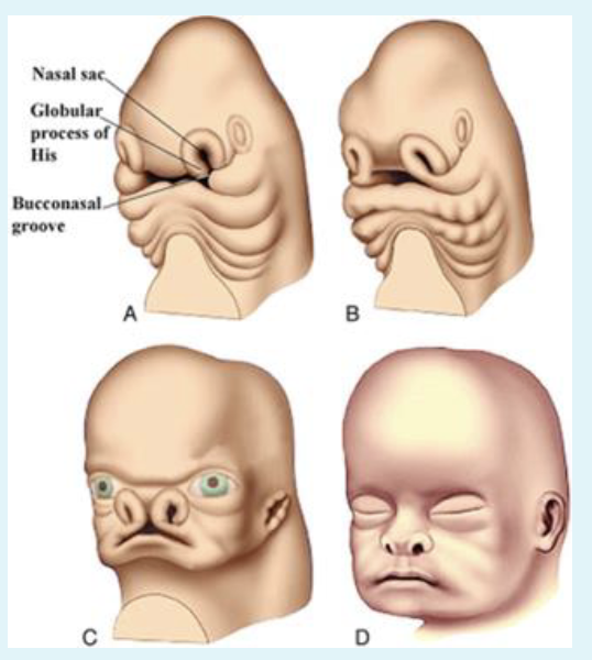
Development of head and face
- Pharyngeal arches and pouches