Crystal arthritis
LO: Compare and contrast the management of septic arthritis and crystal arthritis
Crystal arthritis
- Monosodium urate (uric acid): gout
- Calcium pyrophosphate: pseudogout
| Knee | Volume (ml) | Viscosity | Colour | WBC/mm3 | PMN (%) | Crystals |
|---|---|---|---|---|---|---|
| Normal | <3.5 | Very high | Clear | 200 | <25 | None |
| Crystal synovitis | >3.5 | Low | Straw/opaque | >10,000 | >50% | Present |
Pathogenic process
- Overproduction of uric acid (exo/endogenous)
- Underexcretion of uric acid
- Combination of both
- Most patients with hyperuricemia and primary gout (90%) are underexcretors of uric acid
Drugs causing hyperuricemia
- Cyclosporine
- Alcohol
- Nicotinic acid
- Thiazides
- Lasix (Frusemide)
- Ethambutol
- Aspirin (low dose)
- Pyrazinamide
CANT LEAP
Epidemiology
- Common inflammatory arthritis in men over 40
- After menopause increase in women - loss of uricosuric effect of oestrogen
- Aging population
- Comorbidities
Predisposing factors
- Immediate postop period after major surgery
- Acute myocardial infarction
- Stroke
- Fasting
- Alcohol abuse
- Large intake of food with high purine content
- Local infection
- Kidney stones
Management
- Patient education
- Diet - low purine

- Reduce alcohol
- Weight reduction
- Colchicine prophylaxis for the first 3-6 months to reuce frequency of heart attacks
- Offer urate lowering therapy - lifelong treatment
- Chronic kidney disease stages 3 to 5
- Diuretic therapy
- Tophi
- Chronic gouty arthritis
Acute attack
- Joint rest and local ice
- NSAIDS/COX II
- Oral steroids
- Local steroid injection
- Oral colchicine
Intercritical gout
- Diet
- Alcohol
- Colchicine prophylaxis
- Urate lowering drugs
- 2 or more heart attacks in a year
Pseudogout
- Deposition of calcium pyrophosphate dihydrate (CPPD)
- Chondrocalcinosis - calcified cartilage on x-rays
- Presence of CPPD crystals associated with aggressive, destructive osteoarthritis
Conditions predisposing
- Age
- Hyperparathyroidism
- Hemachromatosis
- Hypomagnesemia
- Hypophosphatasia
- Acromegaly
- Hypothyroidism
- Trauma
- Infection
- Osteoarthritis
Acute pseudogout (a/c)
- 25%
- Elderly women > men
- Knee most involved
- Ankles, wrist and shoulders
- Attacks self limiting
- Surgical procedures
Chronic pseudogout (c/c)
- Pseudo osteoarthritis (OA) or pseudo rheumatoid arthritis (RA)
- Pseudo OA commonest form of c/c CPPD
- Primary OA pattern of involvement of wrist and metacarpophalangeal joint and atypical knee OA
- CPPD crystals associated with more severe OA
- Found in 60% of knee joints at time of replacement
Diagnosis
- History
- Clinical examination
- Joint fluid analysis
- X-ray
| Osteoarthritis | Rheumatoid arthritis | Calcium pyrophosphate dihydrate |
|---|---|---|
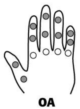 | 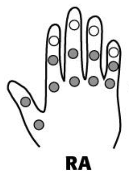 | 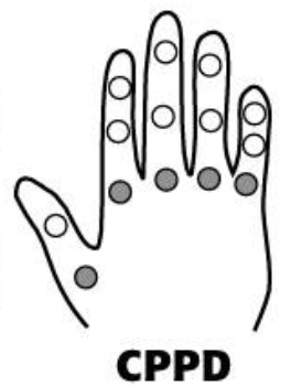 |
White - affected Black - not affected |
LO: Discuss the differential diagnosis of presentations of a/c and c/c monoarthropathy
Aspiration of synovial fluid
- Joint fluid analysis
- White cell count in joint fluid neutrophils
- Culture to rule out infection
- Sodium uric acid
- Renal function
- Urine dipstick - haematuria
Radiography
- Well defined erosions
- Sclerotic borders
- Overhanging edges
- No osteoporosis
- Joint space: symmetric loss vs preservation
- Erosions: marginal vs eccentric with overhanging edge
Differential diagnosis
- Degree of inflammation - different from rheumatoid arthritis
- Matched by other crystal disease (pseudogout) or infection
- Podagra (1st metatarsophalangeal joint pain) characteristic of gout
- Degree of inflammation matched by gout and septic arthritis
- Gout - 1st metatarsophalangeal joint
Treatment
- Acute attack joint aspiration and steroid injection
- Anti-inflammatories
- Chronic pseudogout
- Periodic intraarticular steroid injection
- Associated disease managed
Allopurinol
- Most commonly used drug to lower uric acid
- Xanthine oxidase inhibitor
- Lower serum and uinary uric acid excretion
Gout flare
- Measure serum urate level
- Information about gout, self-management and reduce the risk of future flares
- Assess lifestyle and comorbidities
- Review medications and discuss the risks and benefits of long term urate-lowering therapy
Radiographic appearance
- Calcium deposition
- Triangular fibrocartilage
- Subchondral sclerosis
- Joint space narrowing
- Subchondral cyst formations