Lower limb 1
LO: Identify the muscles of the anterior thigh, medial thigh and anterior leg, and describe their action/innervation
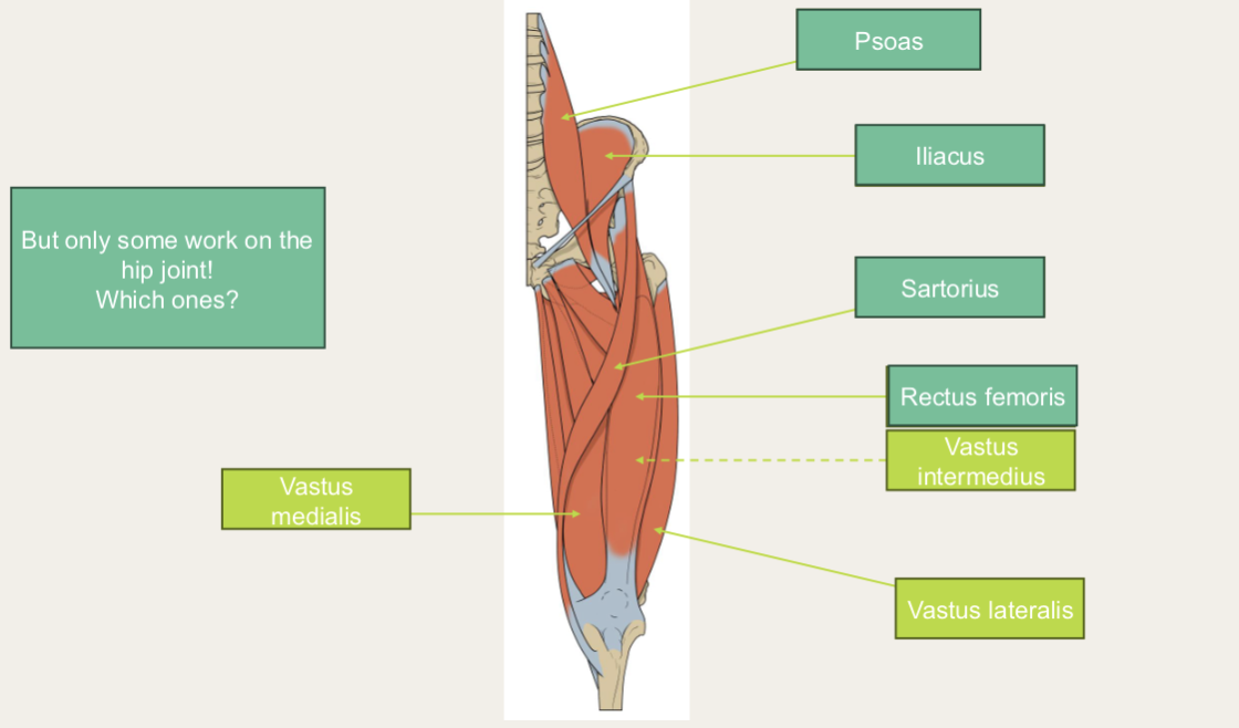
Anterior compartment
- Knee extension
- Femoral nerve
- Femoral artery and vein
- Psoas
- Iliacus
- Sartorius
- Rectus femoris
- Vastus intermedius/medialis/lateralis
Medial thigh
- Innervation: Obturator nerve
- Blood supply: Obturator artery
- Obturator externus
- Pectineus
- Gracilis
- Adductor longus/brevis/magnus
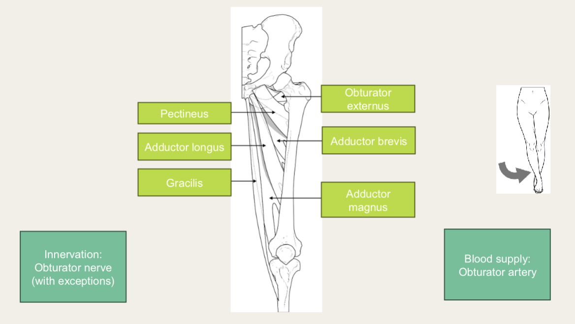
- Adduction
LO: Identify the borders and contents of the femoral triangle
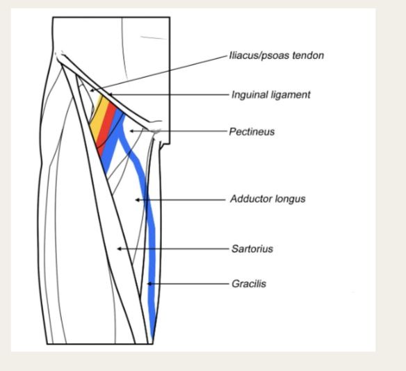
Femoral triangle
- Iliacus/psoas tendon
- Inguinal ligament
- Pectineus
- Adductor longus
- Sartorius
- Gracilis
LO: Identify the three gluteal muscles and describe their action and innervation. Explain the role of gluteus medius/minimus in normal locomotion and relate this to Trendelenburg gait
Gluteal region
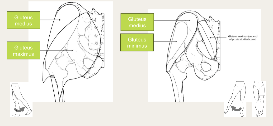
- Gluteus medius/maximus/minimus
-
Blood supply: superior gluteal artery (Gluteus maximus has a supply from inferior as well)
-
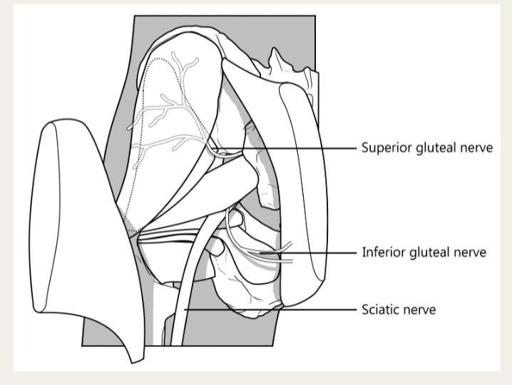
Superior/inferior gluteal nerve
-
Sciatic nerve
-
Extension
-
Abduction
-
Medial rotation
LO: Identify the muscles of the gluteal region that form the deep/lateral rotators and, as a group, describe their function
Deep gluteal muscles
- Innervation: nerves from lumbosacral plexus
- Blood supply: inferior gluteal artery
- Piriformis
- Superior/inferior gemullus
- Obturator internus
- Quadratus femoris
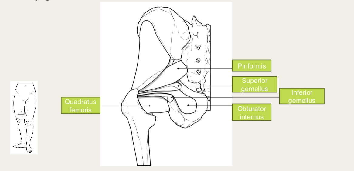
- Lateral rotation
LO: Identify the muscles of the posterior thigh and describe their action and innervation. Explain their role in limiting movements of the hip and knee joints
Posterior thigh muscles
- Innervation: sciatic nerve
- Blood supply: Femoral/popliteal artery
- Biceps femoris (short and long head)
- Semitendinosus
- Semimembranosus
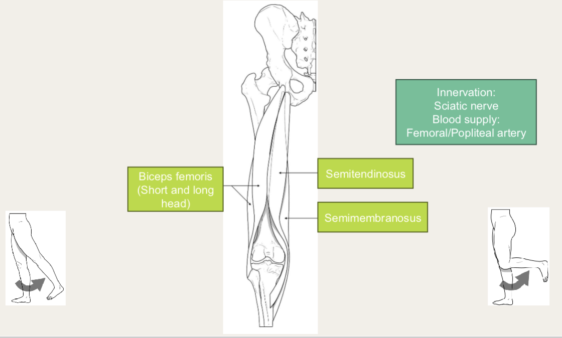
- Extension
- Flexion
LO: Describe which bones form the hip joint and identify the major bony landmarks of the hip, including radiological appearance
Hip
- Ilium - top
- Sacrum - between each hip bone
- Femur attaches at acetabular fossa (acetabulum)
- Ischium - bottom posterior
- Pubis - Bottom anterior
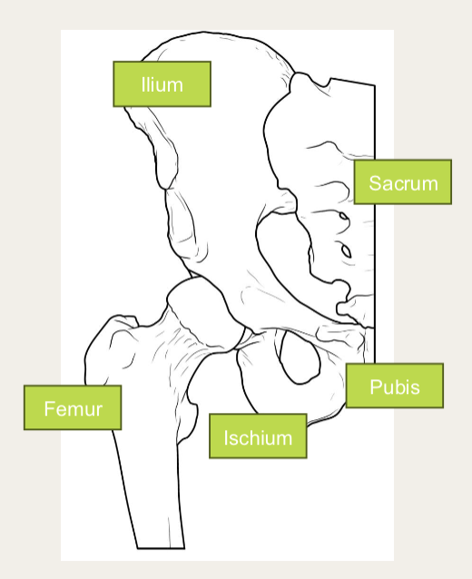
Pelvis
- Anterior superior/inferior iliac spine
- Femoral head/neck
- Greater/lesser trochanter
- Pelvic brim
- Ischial tuberosity
- Iliac crest - top
- Ischial spine
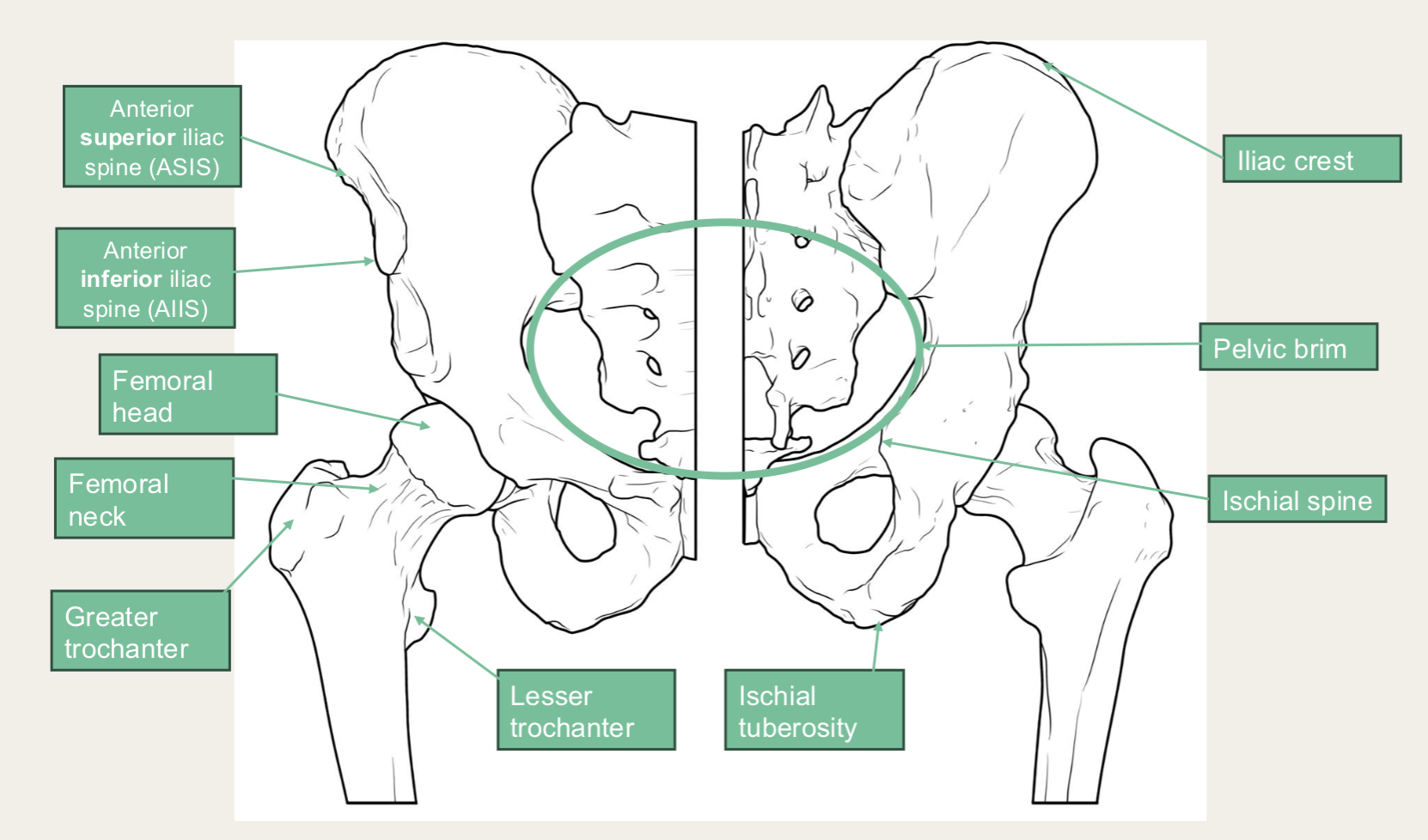
- Fovea - attachment for ligamentum teres
- Greater/lesser sciatic notch
- Acetabular articular surface and labrum
- Ligamentum teres
- Ischial spine
- Transverse acetabular ligament and notch
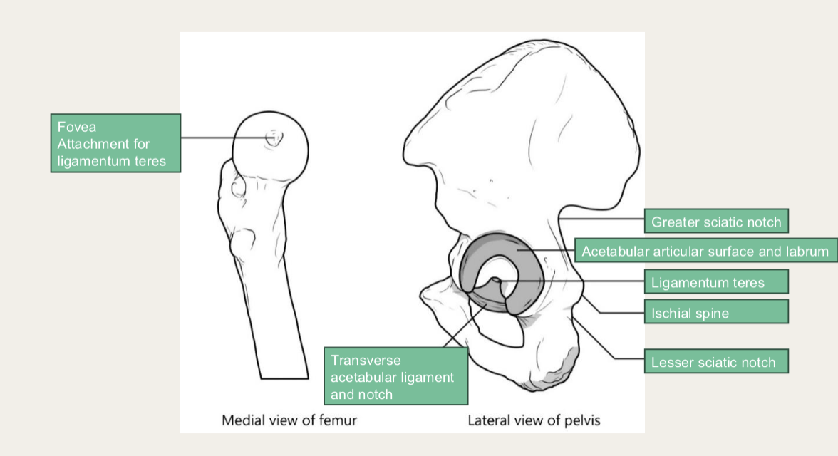
LO: Describe the six movements of the hip joint, the structures that provide support to the hip (bones, ligaments and cartilage) and the features that limit movement
Hip joint
- Synovial joint
- Ball and socket
- Flexion/extension
- Abduction/adduction
- Lateral/medial rotation
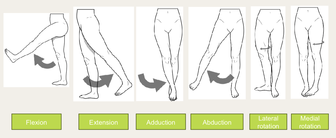
LO: Describe the blood supply to the femoral head at different ages and relate this to neck of femur (NOF) fractures and different treatment options
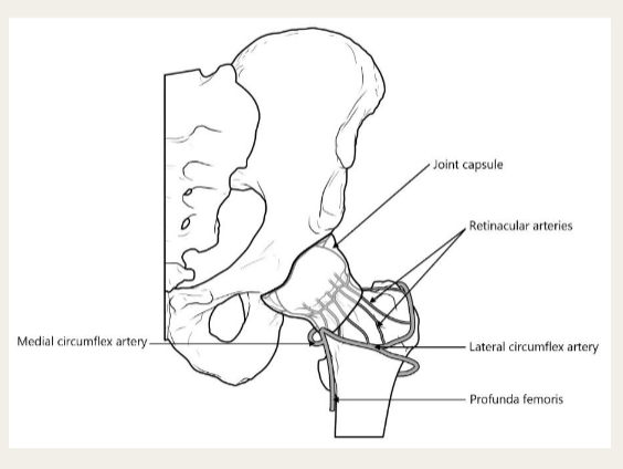
- Obturator artery - through obturator foramen
- Retinacular arteries
- Lateral/medial circumflex artery
- Profunda femoris
LO: Compare and contrast the features of the shoulder and hip joints (bones, ligaments, capsule and cartilage) and consider how they are adapted for function
Ligaments of the hip
- Pubofemoral - pubic to femur
- Iliofemoral - ileum to femur
- Ischiofemoral - ischium to femur
- Ligaments spiral around hip joint
- Flexion - unspirals, loosens
- Extension - spirals, tightens
| Feature | Hip | Shoulder |
|---|---|---|
| Depth of socket | Deep | Shallow |
| Relative size of head to socket | Well matched | Larger humeral head |
| Capsule | Thick and strong | Thin and weak |
| Extra-capsular ligaments | Tight and strong | Loose and weak |
| Mobility/Stability | Stability | Mobility |
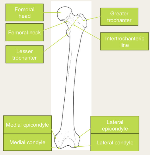
Anterior femur
- Femoral head/neck
- Greater/lower trochanter
- Intertrochanteric line
- Medial/lateral epicondyle
- Medial/lateral condyle
Posterior femur
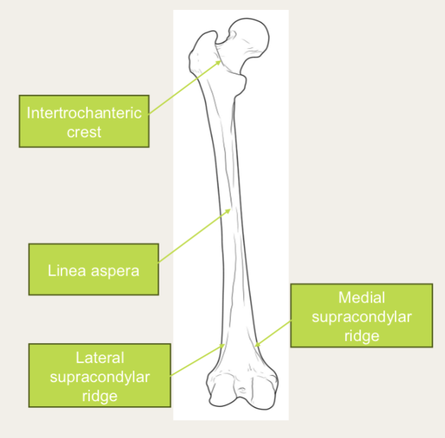
- Intertrochanteric crest
- Linea aspera
- Lateral/medial supracondylar ridge