X-rays
- Beams generated by x-ray tube
- Electromagnetic radiation
- Generator produces x-rays
- X-rays pass through body
- Depends on density of material, denser means less x-rays penetrate so lighter image
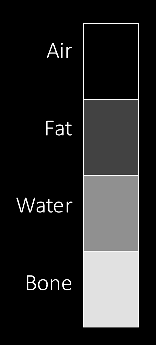
Don't x-ray
Identifying fractures
- Need two views, 90 to each other (orthogonal)
Radiographic signs of fracture
| Bone | Supplementary |
| Lucent lines (dark) | Haemarthrosis (blood in joint, joint effusion) |
| Sclerotic lines (light) | Lipohaemarthrosis |
| Corticol breach | Soft tissue swelling |
| Disruption of trabeculae | |
| Change in bony contour | |
Ring facture
- Look for another fracture
- Ring must break in at least 2 places
- Pelvis - obturator foramen
- Paired long bones
- Mandible
- Zygomatic arch
- Vertebra
Description of fractures
Position
- Angulation
- Displacement
- Distraction
- Impaction
- Foreshortening/shortening
- Position of distal relative to proximal
Path of fracture line
- Transverse
- Oblique
- Spiral
- Longitudinal
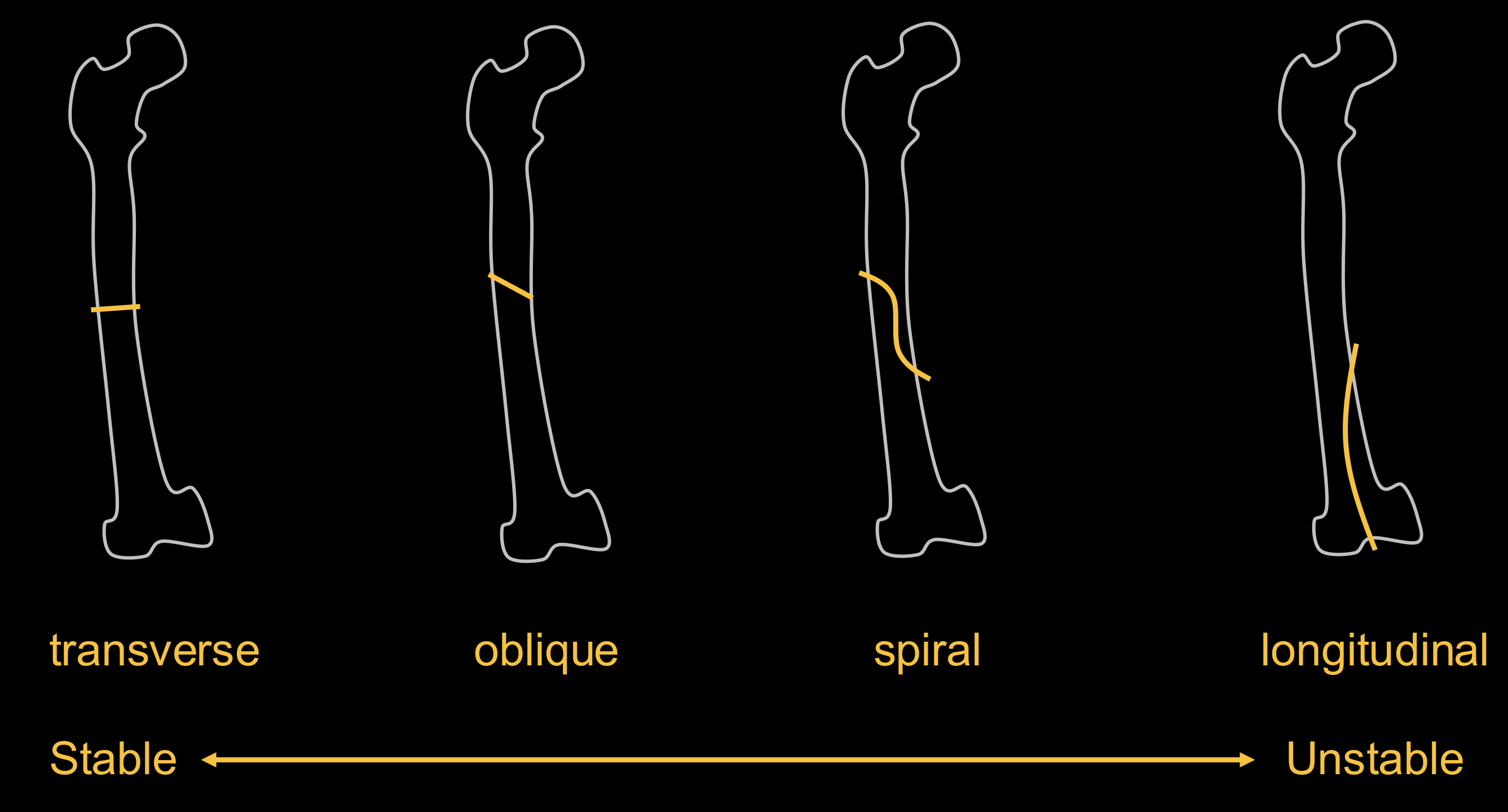
- Verticality of fracture = instability
Simple or comminuted
- Simple - 2 pieces
- Comminuted - 3 or more fragments
Joint involvement
Closed or open (compound)
- Closed - fracture does not pierce skin
- Open (compound) - fracture pierces skin
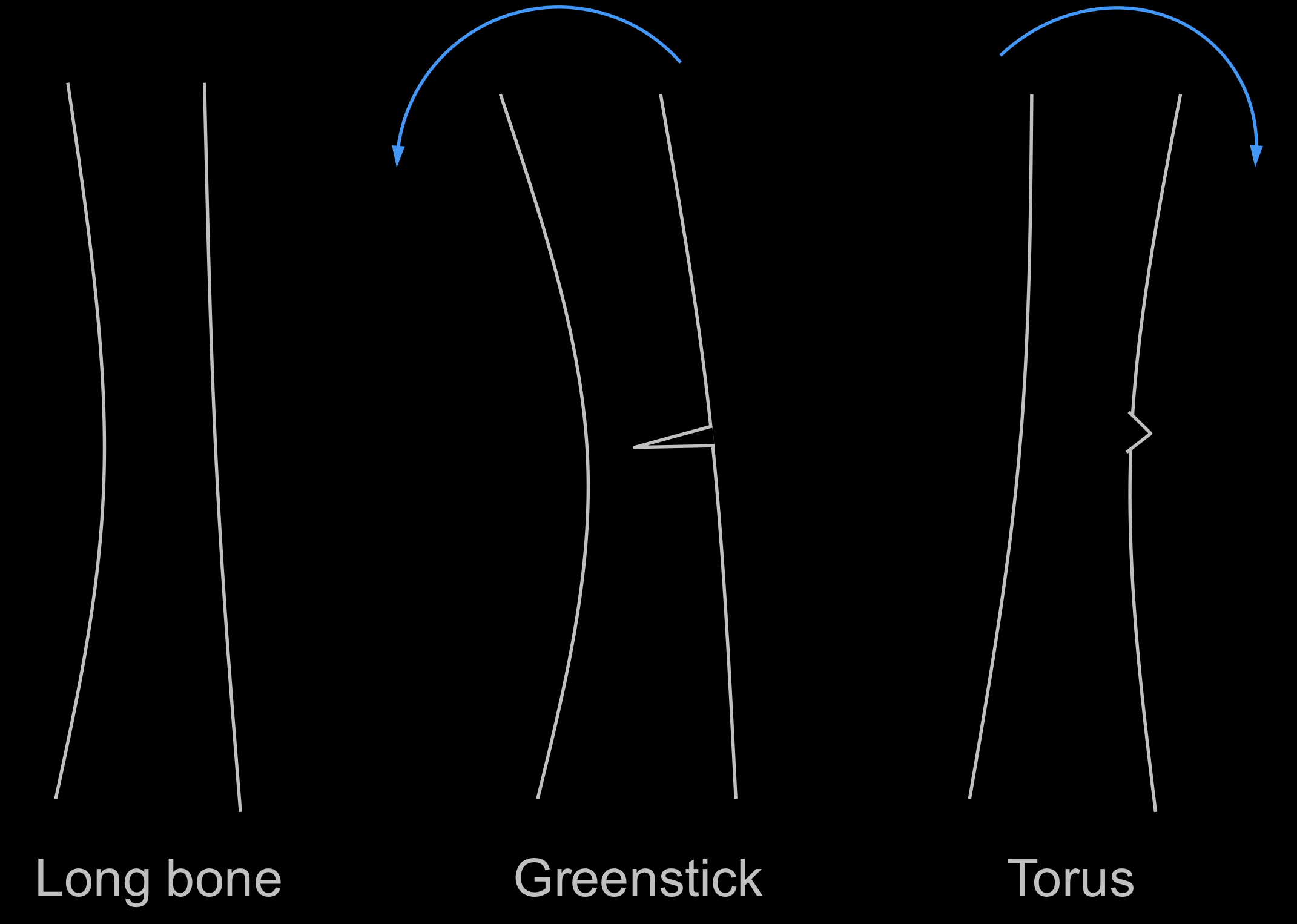
Fractures in children
- Plastic
- Torus (donut/ring)
- Greenstick
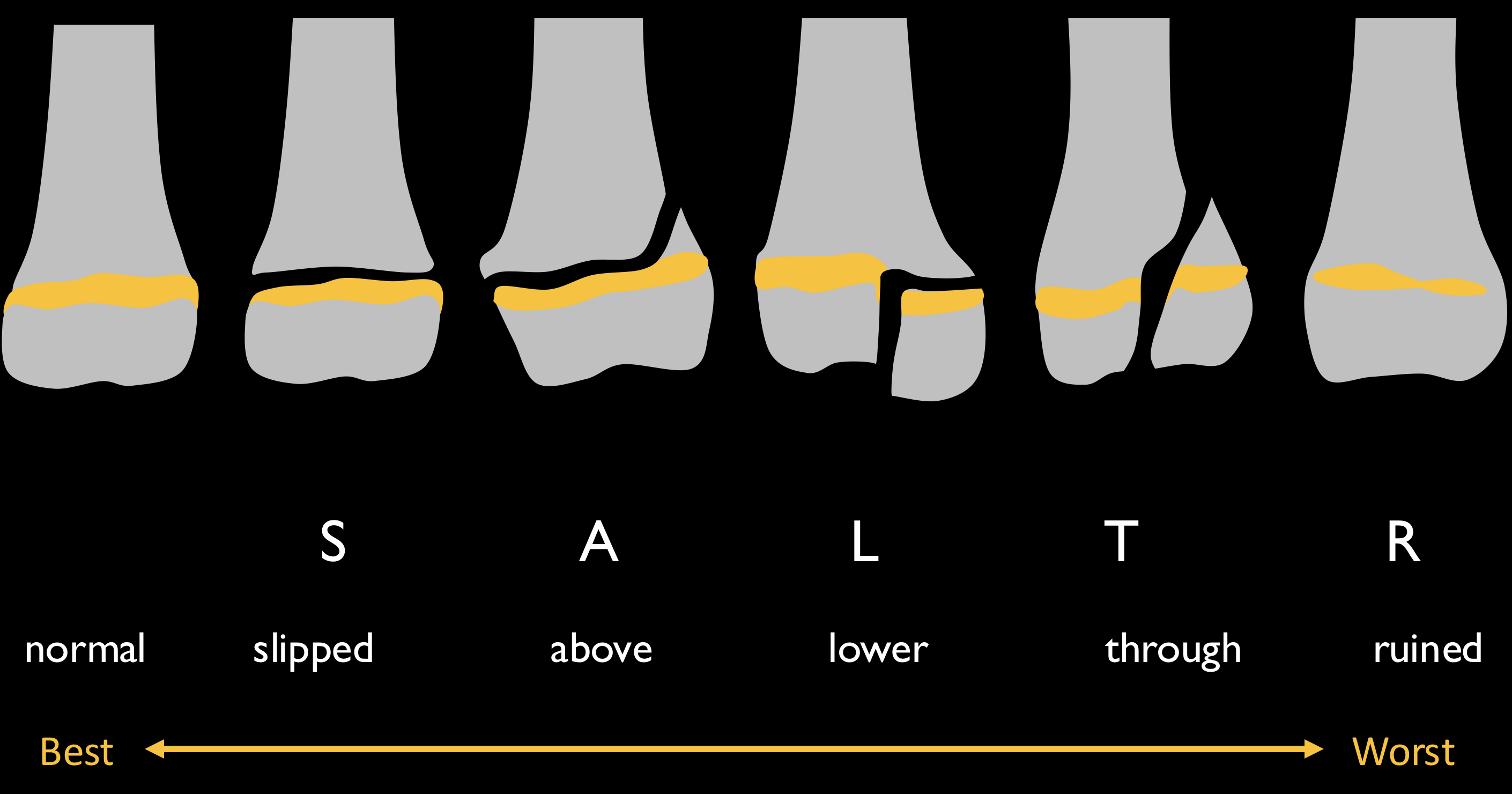
- Growth plate fractures
Salter-Harris classification
Special fractures
flowchart TD
	Fracture --- A[Normal load]
	Fracture --- B[Abnormal load]
	A --- C[Abnormal bone]
	C --- Focal
	C --- Diffuse
	Focal --- Pathological
	Diffuse --- Insufficiency
	B --- D[Normal bone]
	D --- Stress